Key Points:
- The posteromedial bow of the tibia leads to foot and ankle deformities and tibial malformations.
- Often the bowing is spontaneously corrected, but incompletely, in the first four years of life.
- A constant level of growth inhibition is maintained for the tibia
- It is crucial to address leg length discrepancy if it is anticipated that it will exceed 2 cm at skeletal maturity
- Without treatment, the average difference between leg lengths is 3.5 cm
- It is best to delay operative treatment until closer to skeletal maturity unless severe angular deformity cannot be corrected by growth
Description:
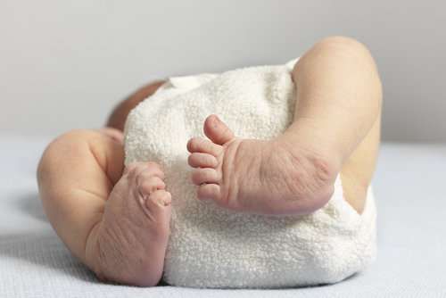
Congenital anomaly in which at least one tibia and fibula are bowing posteriorly and medially. This form of deformity is evident at birth. As a result, the foot assumes a calcaneovalgus position with an extreme amount of dorsiflexion. Occasionally, the foot will be abutting the open tibia during delivery, along with a dimple in the skin over the bow’s apex. The tibia maintains its growth inhibition.
Clinical Findings:
Usually, patients with this condition present at birth with a notable leg deformity. Only one side is abnormal. When resting against the tibial shaft, the foot is customarily dorsiflexed. Contracture of the dorsal structures of the leg and foot will allow the patient to lack normal passive plantarflexion of the ankle.
Additionally, underdeveloped calf muscles and reduced foot size and range of motion are also associated with this condition. A research study also identified ankle valgus as related to this condition. There has been speculation that this might result from a distal fibular physis located proximally or due to the distal tibia physis wedged between it and the fibula.
The posteromedial bow of the tibia will improve with growth, but the most dramatic improvement occurs during the first year of life. Wolf’s law indicates that remodelling at the diaphysis through reorientation results in more significant correction than remodelling at the physis via reorientation. Throughout life, the growth inhibition in the tibia remains constant. Therefore, it is possible to predict the leg length discrepancy at maturity from x-rays acquired at a young age.
The medial bow is related to leg length discrepancy. It is found that the greater the bow, the longer the leg is. A growth inhibition percentage ranging from 15-40% also results in an average discrepancy of 3.5 cm.
Study of images:
Initial imaging studies to evaluate the condition are AP and lateral tibia radiographs. A tibial bowing improves with growth, but a leg length discrepancy persists because of growth inhibition. Periodic leg length radiographs should be conducted to monitor the condition.
Etiology:
We do not know what causes posteromedial bowing. Several theories explain the phenomenon, including mechanical, e.g., the dorsiflexed foot hitting the tibia or abnormal placentas (amnion rupture).
Treatment:
A stretching program for the dorsal structures of the ankle is the first treatment for an infant with this condition. Once parents have been instructed appropriately, these stretches are performed at home. During infancy, children with severe contractures may require serial casting.
Treatment focuses on managing residual bowing and leg length discrepancy due to the resulting leg length discrepancy. There have been many methods of treatment proposed with differing outcomes.
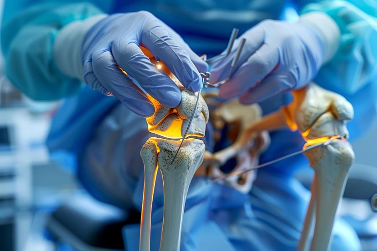
Leg length discrepancies can be treated either by contralateral epiphysiodesis or a frame to lengthen the leg. Osteotomy with external fixation has the advantage of correcting any residual angular deformity.
According to recent studies, since the final angular deformities with and without surgery do not differ at maturity, delaying surgery until skeletal maturity can provide a more accurate correction of the leg length discrepancy and increase the patient’s compliance with their care.
Complications:
While the ankle range of motion has not been altered following surgery, its arc remains unchanged. A surgical procedure to correct leg length discrepancies can cause complications such as pin tract infections or non-union. The long-term need for orthosis and the cost are the main drawbacks of nonoperative therapy. The difference in leg length greater than 2 cm can affect the development of back, hip, or knee arthritis in the future.
Conclusion
During the first few years of life, posteromedial bowing of the tibia gradually improves, but limb reconstruction is indicated in 20/38 (53%) cases where significant residual deformity or progressively worsening LLD are present. Treatment options include limb reconstruction with a hexapod external fixator as a treatment option for larger discrepancies and persistent deformities.