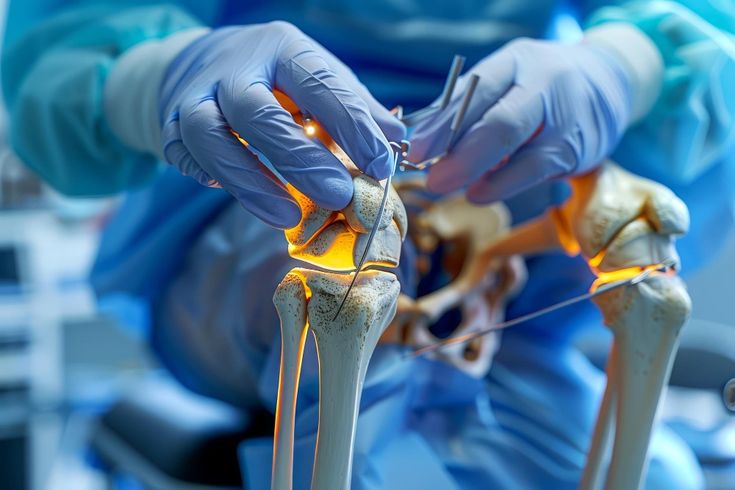
Lesions in the bone are masses of tissue that can develop in any part of the bone.
It is possible to develop bone lesions in any portion of the body and any piece of bone, ranging from the surface of the bone to the interior of the bone marrow.
They are caused by uncontrolled cell division and proliferation in the bone, resulting in a lump or mass of abnormal tissue.
A growing lesion can erode healthy tissue and weaken the bone, increasing its susceptibility to fractures.
The majority of bone lesions are benign, do not pose a threat to the patient's life, and do not spread to other areas of the body.
However, certain bone lesions are malignant, or cancerous. These bone lesions can occasionally metastasize, which refers to the spread of cancer cells to other areas of the body.
Two distinct types of malignant bone tumors exist:
- Cancer that begins in the bone is referred to as primary bone cancer.
- Secondary bone cancer occurs when cancer spreads from another location to the bone.
The cause of a bone lesion is determined by its benign or malignant nature, as well as other factors.
Benign bone lesions
The majority of bone lesions are benign, which means they are not cancerous or life-threatening. Additionally, certain diseases and conditions resemble bone lesions. Benign bone lesions are caused by a variety of factors, including the following:
● Fibromatosis that is not ossifying
● Bone cyst with a unicameral (simple) structure
● Osteochondroma
● tumor with giant cells
● Enchondroma
● Dysplasia of the fibrous connective tissue
● Chondroblastoma
● Bone cyst with aneurysm
● Osteoid osteoma is a type of osteoma.
If the lesion is benign, your doctor may recommend periodic X-ray monitoring. Certain lesions, particularly those in children, may resolve over time.
Medicines may successfully treat other types of bone lesions. In some instances, surgical removal of the lesion may be necessary to avoid a bone fracture.
Benign lesions may recur following treatment. They may spread or become malignant in rare instances.
Malignant bone tumors
There are two types of cancer: primary cancer and secondary cancer. The cause of malignant bone tumors or cancerous bone lesions depends on which type of cancer it is.
Primary bone cancer lesions are caused by a variety of factors, including:
Multiple myeloma
Multiple Myeloma is the most common form of primary bone cancer in people over the age of 50. Multiple myeloma is a malignant tumor of the bone marrow, the soft tissue located in the center of bones that produces blood cells. There are approximately six people per 100,000 every year who are affected by primary bone cancer every year. It can affect any bone in the body and is the most frequently occurring primary bone cancer. Between the ages of 50 and 70, the majority of people develop multiple myeloma. chemotherapy and radiation therapy are usually used to treat multiple myeloma. Surgery may be necessary on occasion. Multiple myeloma has a 5-year 5-year survival rate of 50 percent. That means that slightly less than half of those diagnosed with the disease will survive five years after diagnosis.<>
Osteosarcoma
Osteosarcoma is the second most frequently occurring primary bone cancer after osteoporosis. It is still uncommon, affecting between two and five people in every million each year. The majority of osteosarcoma cases occur on either side of the knee in teenagers and children's thigh bone or shinbone. Additionally, it can occur in the hip or shoulder on rare occasions. Chemotherapy and surgery are usually required for treatment. Children and adolescents with osteosarcoma in a single location have a 5-year survival rate of 70 percent. Chemotherapy, surgery, and radiation therapy are typically used to treat osteosarcoma.
Ewing sarcoma
Ewing sarcoma is most likely diagnosed in children and adolescents between the ages of 5 and 20. This type of tumor most commonly affects the upper and lower legs, pelvis, upper arm, or ribs. Additionally, it can develop in the soft tissue that surrounds a bone. While Ewing sarcoma can occur at any age, it affects more than half of those diagnosed between the ages of 10 and 20. Overall, the 5-year survival rate for children and adolescents with unresectable Ewing sarcoma is approximately 70 percent. If the tumor has already spread when diagnosed, the prognosis is less favorable. Chondrosarcoma
Chondrosarcoma is a malignant tumor consisting of cartilage-producing cells. It is mainly seen in adults between the ages of 40 and 70. In the hip, pelvic, or shoulder area, these tumors tend to grow. Chondrosarcoma is commonly treated with surgery but depends on the stage and severity of the malignancy. During limb-sparing operation, a metal replacement or bone graft is used to remove the damaged section of the bone. Occasionally, the area affected may need to be removed if the cancer cells have progressed from the bone into nerves and blood vessels.
Chondrosarcoma is a slow-growing malignancy, with most instances being diagnosed at low levels.
Secondary bone cancer lesions Oncology specialists have identified several types of cancer that start elsewhere in the body and can move to the bone. These include:
● breast
● lung
● thyroid
● renal
● prostate
If you have secondary bone cancer that has migrated from another site, the treatment options and prognosis will be determined by the type and severity of your underlying cancer.
Dull pain, stiffness, and swelling in the affected area are some indications of bone lesions.
Occasionally, bone lesions might result in localized pain. This type of pain is typically dull or painful and may become worse with movement. Additionally, a fever and nocturnal sweats may occur.
Along with discomfort, certain malignant bone lesions might result in stiffness, edema, or sensitivity in the affected area. The pain may fluctuate and may be more severe or less severe at night.
Not everyone will have these symptoms; instead, they may discover a non-painful lump on their body.
Lesions in the bone can deteriorate the structure of the bone, making it more susceptible to fracture. As a result, an individual who has a bone lesion may break a bone without being injured.
A doctor will do a thorough physical examination as well as several tests to determine the source of a bone lesion. They may inquire about your overall health, medications, and symptoms, as well as whether you have a family history of lesions or cancer.
At the time of your physical examination, a doctor will examine for swelling or pain, any changes in your skin, the existence of a mass, and whether or not the condition has an impact on your joints.
Aside from that, they will request imaging tests, which might include X-rays, magnetic resonance imaging (MRI) scans, and computed tomography (CT) scans among other things.
A biopsy may also be required to achieve a definitive diagnosis. During a biopsy, a small sample of the lesion will be taken and examined under a microscope for possible treatment options. Further testing may include the taking of blood and urine samples.
Osteomyelitis complications may include:
• Bone death (osteonecrosis). An infection in your bone can impede blood circulation within the bone, leading to bone death. Areas where the bone has died need to be surgically removed for antibiotics to be effective.
• Septic arthritis. Sometimes, infection within bones can spread into a nearby joint.
• Impaired growth. Normal growth in bones or joints in children may be affected if osteomyelitis occurs in the softer areas, called growth plates, at either end of the long bones of the arms and legs.
• Skin cancer. If your osteomyelitis has resulted in an open sore that is draining pus, the surrounding skin is at higher risk of developing squamous cell cancer.
If you've been told that you have an increased risk of infection, talk to your doctor about ways to prevent infections from occurring. Reducing your risk of infection will also help your risk of developing osteomyelitis.
In general, take precautions to avoid cuts, scrapes and animal scratches or bites, which give germs easy access to your body. If you or your child has a minor injury, clean the area immediately and apply a clean bandage. Check wounds frequently for signs of infection.
The prognosis for individuals who have a bone lesion is determined by the type of lesion.
Benign lesions may require only close observation or pharmacological treatment, though they may recur following effective treatment.
Following therapy, individuals with malignant bone lesions will need to contact their doctor frequently, typically every few months, to monitor for symptoms of recurrence.