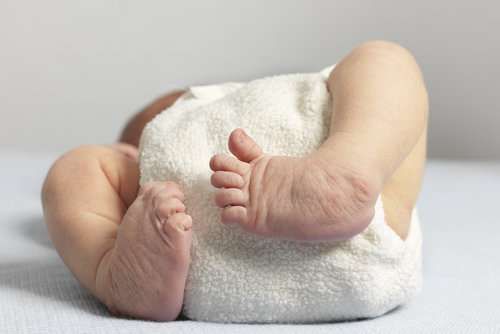
What Is Erb’s Palsy?
Erbs Palsy is a group of neurologic injuries of the brachial plexus observed in newborns. Erb and Duchenne separately described neurologic injuries to C5 and C6 nerve roots that are now collectively named Duchenne-Erb palsy while Klumpke described injury to C8 and T1 nerve roots. (Duchenne 1872; Erb 1874; Klumpke 1885)
Mechanisms of Erb’s Palsy
One of the most common causes of Erb’s palsy is a condition called shoulder dystocia, which occurs when an infant’s shoulder is caught behind the mother’s pubic bone during childbirth. When a medical professional pulls on the baby to release their shoulder, it can stretch or tear the healthy nerves in that area.
Other situations that may cause Erb’s palsy:
- The baby’s head and neck pulling sideways as they pass through the birth canal
- Pulling the baby’s shoulders during a head-first delivery
- Pulling on the baby’s feet during a feet-first (breech) delivery, which puts too much pressure on the infant’s arms
- Shoulder dislocation or fractures in the collarbone (clavicle)
Description:
Neonatal brachial plexus palsies (NBPP) Involvement ranges from mild weakness to a flail extremity. Many patients, but not all, will experience partial to full spontaneous recovery in the first months of life.
Clinical Findings:
Narakas described four separate groups of palsies:
- Group I is the classic Erb’s palsy, C5-C6, with absence of shoulder abduction and external rotation, elbow flexion, and forearm supination with intact wrist and finger flexion and extension.
- Group II involves C5-C7, with absence of wrist and finger extension in addition to the impairments described above, leading to the classic “waiter’s tip position.”
- Group III involves C5-T1 and results in a flail extremity but without an associated Horner’s syndrome.
- Group IV includes not only a flail extremity with C5-T1 involvement but also an associated Horner’s syndrome. (Narakas, 1987)
Associated clinical findings that increase suspicion about pre-ganglionic lesions include
- Horner’s syndrome;
- elevated hemidiaphragm from phrenic nerve involvement;
- winged scapula from long thoracic nerve involvement;
- and the absence of rhomboid, rotator cuff, or latissimus dorsi function.
- Other possible injuries in neonates with NBPP include fractures of the clavicle and humerus, shoulder subluxation, cervical spine subluxation, cervical spinal cord injuries, and facial palsies.NBPP is diagnosed during the newborn physical exam with findings of weakness and/or limited arm movements. It is important to look for asymmetric reflexes in the upper extremities as well as the other findings of lower and upper trunk involvement (e.g. ptosis, miosis, winged scapula, and asymmetric chest expansion). If there is muscle atrophy or muscle contracture leading to restricted passive range of motion on newborn exam, clinical suspicion should be high for in-utero nerve injury rather than injury during delivery due to the time it takes to develop these findings. (Hoeksma, 2000) The Active Movement Scale (AMS) assists in the evaluation of newborns and can document functional muscle recovery.
In older children, brachial plexus function is commonly measured using the Mallet classification system, which incrementally evaluates a patient’s global abduction, global external rotation, and movements from hand to neck, hand to spine, and hand to mouth. (Mallet 1972)
Patients with NBPP may develop internal rotation contractures of the shoulder (50-70%), glenohumeral dysplasia, or dislocation (8%) due to a lack of active external rotation and muscle imbalance.
Imaging Studies:
Radiographs of the clavicle and humerus are useful to evaluate for potential fractures if there is concern for NBPP.
Further imaging such as MRI and CT myelogram can investigate the cervical spinal cord, nerve roots, and brachial plexus to localize the injury.
Electrodiagnostic studies may also be useful in evaluation and localization of the injury. Vanderhave et al found electrodiagnostic studies to be more sensitive for upper trunk ruptures (94.6%), while CT myelogram was more sensitive for lower trunk avulsions (83.3% at C7 and 75.0% at C8-T1). (Vanderhave, 2012)
Electrodiagnosis (EDX) is a useful test to accurately localize the site, determine the extent, identify the predominant pathophysiology, and objectively quantify the severity of brachial plexopathies. It can also be used to examine muscles not easily assessed clinically and recognize minimal defects. Post-operatively and on follow up studies, it is important for early detection of re-innervation Localization of the site of the lesion can be very challenging as there may be multiple sites of involvement and hence the electroneuromyographic evaluation must be adequate. The unaffected limb also needs to be examined for comparison. The final impression must be co-related with the type and severity of injury.
Etiology:
Although the most common mechanism of NBPP is believed to be stretch injury, others include nerve compression, nerve infiltration, and oxygen deprivation. These injury mechanisms may occur simultaneously, such as shoulder dystocia leading to nerve stretch injury in conjunction with oxygen deprivation due to compression of the umbilical cord. While NBPP has traditionally been attributed to an iatrogenic, lateral traction force during shoulder dystocia, it may also occur following an uncomplicated vaginal delivery or cesarean section. (Al-Qattan, 1996) Risk factors for NBPP include shoulder dystocia, excessive maternal weight gain, maternal diabetes, multiparity, fetal macrosomia, fetal malposition, labor induction, prolonged labor, operative vaginal delivery, and prior shoulder dystocia or NBPP pregnancies. (Gherman, 2014, Lindell-Iwan 1996)
Types of Erb’s Palsy Injuries
The severity of a child’s condition depends on the type of nerve damage that occurs:
- Neurapraxia:Stretching of the nerve without tearing
- Neuroma:A stretch injury that may cause scar tissue
- Rupture:Tearing of the nerve without separation from the spinal cord
- Avulsion:The nerve root tears away from the spinal cord and will not heal on its own
Treatment:
The majority of patients with NBPP will recover spontaneously, and those that recover antigravity upper strength of muscles innervated by the upper trunk by 2 months of age will regain full function by age 1-2 years. (Waters, 2005)
Initial treatment involves maintaining passive range of motion with physical therapy and splinting to prevent contracture while awaiting spontaneous recovery. Infants who have not recovered antigravity biceps strength by 5-6 months of age will have permanent limitations and may benefit from surgical treatment.
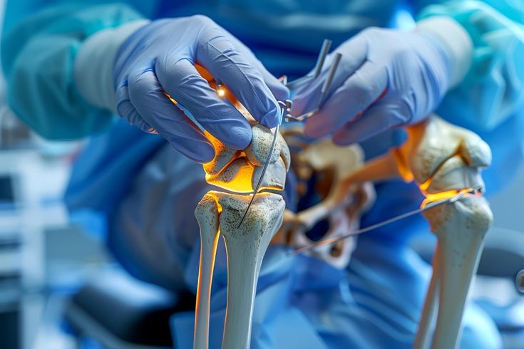
Surgical treatment may directly address the neurologic injury (microsurgery such as neuroma excision and nerve grafting or nerve transfers), or may be aimed at improving function via soft tissue release, tendon transfers, or osteotomy.
Preganglionic lesions are often treated with nerve transfer as they cannot be repaired directly and will not recover spontaneously. Postganglionic ruptures may be treated with neuroma excision and nerve grafting or other reconstruction if functional recovery is not occurring.
There is debate in the literature regarding when to pursue surgery, but many academic centers use the following as definitions for incomplete functional recovery: inability to flex the elbow against gravity by three months of age, impaired or incomplete hand function at three months in a baby born with a flail arm, the towel test (inability to uncover his or her own face at 6 months), and the cookie test (inability to bring cookie to mouth at nine months) Surgery to treat the shoulder may include open or arthroscopic soft tissue releases and muscle transfers to correct internal rotation contracture and to promote glenohumeral remodeling. Open or arthroscopic reduction may address glenohumeral dislocations. Botulinum toxin has also been used to treat contractures. Humeral osteotomy may be used in an older child or adolescent to place the arm in a more functional position.
Complications:
The most common complications are incomplete functional recovery and contractures, particularly shoulder internal rotation due to reduced infraspinatus function. Altered muscular forces on the glenohumeral joint lead to abnormal posterior loading and ultimately a flattening or biconcave glenoid and subsequent shoulder instability. Other common complications include poor active shoulder elevation and scapular dyskinesia.
Key Points:
- The majority of patients with NBPP will recover spontaneously
- Early treatment is focused on prevention of contractures and maintenance of glenohumeral reduction
- Surgical treatment is indicated for preganglionic avulsions and postganglionic injuries not demonstrating return of function
- Infants who have not recovered antigravity biceps strength by 5-6 months of age will have permanent limitations and may benefit from surgical treatment
- Surgical options include early microsurgical treatment of the neurologic injury or later soft tissue balancing, tendon transfers, or osteotomies to improve function