Key Points:
- Congenital knee dislocation (CKD) is a hyperextension deformity of the knee with anterior tibia displacement, present at birth.
- CKD is rare, but is often associated with arthrogryposis, Larsen syndrome, or congenital knee and hip differences. When associated, it is more resistant to non-operative treatment.
- Treatment consists of casting trial as soon as possible; if resistant, several surgical options exist including quadriceps lengthening, soft tissue release and ACL advancement, or femoral shortening.
Description:
Congenital knee dislocation (CKD) is a rare condition that involves hyperextension of the knee joint with varying degrees of anterior tibia displacement diagnosed at birth. The most common classification used defines simple hyperextension (Grade 1), anterior tibial subluxation reducible with flexion (Grade 2), and true anterior tibial dislocation (Grade 3). (Curtis, 1969; Neibauer, 1960)
Epidemiology:
CKD incidence has been reported at 1 in 100,000 live births, or, to better conceptualize its rarity, 1% of the incidence of congenital hip dislocation.
Clinical Findings:
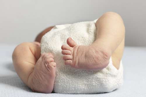
Congenitally dislocated knees have hyperextension contractures with transverse anterior skin folds due to a shortened, sometimes fibrotic quadriceps, tight anterior capsule, and hypoplastic suprapatellar bursa.The tibia may have rotatory or valgus deformity, the hamstrings may displace anteriorly and act as extensors, and there may be general laxity from cruciate pathology or displaced collateral ligaments. Deformity and knee hyperextension can be so severe that the infant’s resting position is with the feet adjacent to the head. Breech presentation in seen in up to 30%, clubfoot in up to 47%, and hip dislocations in up to 50%. CKD has also been commonly associated with arthrogryposis, myelodysplasia, or syndromes such as Larsen, Ehlers-Danlos, or Beals syndrome. Absence of these syndromes and associated deformities, initial range of motion, and time to treatment are the best prognostic indicators.
Imaging Studies:
The diagnosis of CKD is primarily clinical and radiographs are used as a confirmatory study. Radiographs confirm contact between the femoral and tibial epiphysis in Grade 1 and 2 CKD, whereas in Grade 3 CKD the epiphyses are not in contact and the tibia is anterior to the femur. There may be a role for arthrography in knees that fail conservative treatment to better understand the pathologic anatomy prior to surgical intervention, but some dispute its usefulness in guiding management.
Etiology:
Several etiologies have been suggested, and most center on mechanical causes such as decreased intra-uterine space or fetal malposition.Others have suggested quadriceps fibrosis and contracture to be responsible.
Treatment:
Most Grade 1 and 2 CKD can be treated non-operatively, with serial casting with or without supplemental skin traction.Treatment is best when applied as soon as possible, with some advocating initiating treatment less than 20 hours after birth. Once 90 degrees of knee flexion has been achieved, a splint or Pavlik harness can be utilized to maintain correction until the tendency for recurrence has passed. In the presence of a more severe quadriceps contracture, a botulinum toxin injection may assist with progressive stretching and knee flexion.
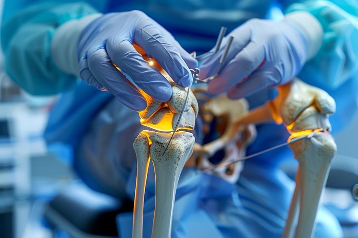
If casting fails, surgery has traditionally consisted of quadriceps lengthening; hamstring, IT band, and capsular release; possible ACL advancement. Recent studies, pointing to long term quadriceps insufficiency with this approach, advocate instead for femoral shortening osteotomy to produce relative lengthening of the quadriceps mechanism. Posterolateral and posteromedial capsulorraphies after excision of redundant posterior capsule may reduce laxity of the knee and the tendency for subluxation. Anterior cruciate reconstruction can be considered if there is excessive anterior laxity at the time of open reduction.
Minimally invasive treatment of CKD has been reported with mini-open and percutaneous quadricepsplasty.
Treatment of CKD prior to treating concomitant hip or foot pathology has been a common practice guideline, but at least one recent study endorses simultaneous treatment of hip and knee dislocation using dual open reductions and femoral shortening osteotomy. Clubfoot casting may also be carried out simultaneously with casting for CKD.
Complications:
In general, CKD treated both operatively and non-operatively have favourable functional results. Closed reduction attempts without first applying traction to clear the tibia distally from the femur has led to epiphyseal deformity. Residual hyperextension may persist in those casted, and laxity or valgus deformity is found in some post-operatively. Quadriceps insufficiency has been associated with open reduction and quadriceps lengthening.