Second-trimester anomaly scan or level-II scan
It is a detailed scan done at 18-23 weeks, during which each part of the fetal anatomy is examined to see if the baby is developing in a normal manner. Particular care is taken towards the brain, face, heart, spine, stomach, bowel, kidneys and limbs.
If any abnormalities are observed, the particular specifications about the findings will be discussed and the couple will be allowed to have further counselling with a Senior Fetal Medicine Consultant.
Why have a Fetal Anomaly Scan?
The vast majority of babies are normal. Nevertheless, all women, have a small possibility of delivering a baby with structural abnormalities that cause physical or mental limitations. Many such abnormalities can be diagnosed and discarded with the help fetal anomaly scan.
Is it safe?
Ultrasound has been used for 40 years to observe pregnancies. So far the data has been convincing that ultrasound is safe for mother and baby. Yet, we believe it is wise to scan only if there is sufficient reason and to use the minimum amount of sound waves based on the ALARA principle – As Low As Reasonably Achievable.
What might be seen?
Most of the major abnormalities can be detected with the help of a scan. However, it is impossible to notice all problems and some will only be found after birth. Conditions like cerebral palsy and autism cannot be observed on a scan.
The position of the baby and the size of the mother are some of the factors that affect the quality of the images. For example, it will be tougher to see the baby clearly if the mother is obese. A poor picture will affect our ability to detect the problems.
During the scan, to study the growth of the baby the doctor measures different parts of the baby’s body. The doctor will measure your baby’s:
- Head circumference (HC)
- Abdominal circumference (AC)
- The femur or thigh bone (FL)
- Humerus or arm bone
In consideration of the anticipated due date, the measurements should match up with the expected values.
The placenta will be reported as low if it touches down to or covers the neck of your uterus (your cervix). If the placenta is low lying, another scan will be scheduled in the eighth month to check the situation once again. By then, it is to be expected that the placenta will have moved away from your cervix.
Later the doctor will observe the baby’s head, face, spine, abdominal wall, heart, stomach, kidneys, umbilical cord, hands, feet, amniotic fluid in detail.
What sort of problems can be found?
Different types of congenital abnormalities and how likely scanning is to identify each problem are mentioned in the table below :

This way that even in case your scan is regular there’s a small danger that your baby will still have a hassle.
Some conditions, consisting of certain coronary heart defects and bowel obstructions, cannot be seen until the 18-20th week of your pregnancy. Having your anomaly scan will probably rule out most of these situations because the giant majority of babies are born healthy.
Can Down syndrome or chromosomal abnormalities be visible on the scan?
This experiment also can pick out 50% to 60% of cases of Down syndrome. Nevertheless, the First Trimester Screening (FTS) check is higher for this. Because 30% to 50% of cases of Down syndrome seem every day on ultrasound, handiest an amniocentesis (checking out the fluid surrounding the infant for its chromosomes) can provide you with these records for sure.
Sometimes infants with chromosomal abnormalities have signs and symptoms known as ultrasound markers. These consist of so as of significance of thick pores and skin behind the neck (nuchal fold), absent nasal bone, moderate fluid inside the ventricles of the mind, an aberrant subclavian artery in the neck, now and again short fingers or legs, white spots within the baby’s coronary heart or abdomen, or choroid plexus cysts in the brain.
While a few infants with chromosomal abnormalities have these markers, it is essential to remember the fact that many everyday babies also have those signs. The handiest way to diagnose or exclude a chromosomal problem for positive is to have an amniocentesis.
Other problems can also suggest that a toddler needs surgical operation or remedy after the start, or even surgical operation while still in the uterus. There will be an entire range of humans to support you through any hard instances, inclusive of obstetricians, paediatricians, pediatric surgeons, pediatric cardiologists/neurologists and so forth.
Which skeletal anomalies are detected commonly?
Clubfoot (CTEV) & Vertebral Malformations (Hemivertebra, Spina Bifida)
An effort is needed to define the character of the limb abnormality with the aid of the following type:
Dysplasia: It is a medical condition in which the cells are arranged into an odd organization within the tissue or an organ. In other words, we can also say it is a method or consequence of histogenesis.
Sequence: A pattern of numerous abnormalities caused by a single known or presumed prior anomaly or mechanical factor
Syndrome: A identified sample of various malformations having one aetiology.
Association: Non-random concurrence of independent malformations, the aetiology of which (one or more) is unknown. This includes the VACTERL association:
Vertebral abnormalities, Anal atresia, Cardiac anomalies, T-E fistula, Esophageal atresia, Renal dysplasia, and Limb/radial irregularities.
Malformation-Deformation: Positional Abnormalities
Positional abnormalities can be considered as defamation or malformation.
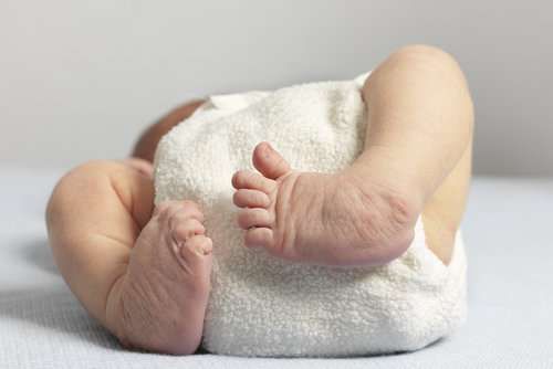
Clubfeet
Clubfoot or talipes equinovarus is a medical condition in which the foot is fixed in adduction, supination, and varus position. There is subluxation of the talocalcaneonavicular joint, with underdevelopment of the tissues towards the midline of the foot and usually of the calf and peroneal muscles. As a result, the foot typically turns inward, and it has a clublike form.
This is one of the most popular congenital birth defects and has been identified as early as 13 weeks gestation by transvaginal sonography and at 16 weeks with a transabdominal ultrasound scan. Approximately one-third of cases are secluded; however, many are linked with other abnormalities such as central nervous system defects and chromosome irregularities. Thus, it is essential to perform a precise fetal ultrasound test, as well as fetal karyotyping.
Clinodactyly
Clinodactyly is a set deviation of the digits. Clinodactyly of the arms can be observed on fetal ultrasound tests, whereas clinodactyly of theft is difficult to detect. This abnormality influences each of the hands, but is normally far visible because of the fifth finger clinodactyly. This abnormality is a result of asymmetrical hypoplasia of the mid-phalanx with the middle portion being shorter than the lateral part, developing radial angulation of the distal phalanx.
Clenched hand
In clenched arms the second one and fifth hands overlap the 0.33 and fourth with an adducted thumb, it is essential to assess, on an ultrasound experiment, if it’s far a chronic or a brief locating. When consistent, it shows the opportunity of chromosomal abnormalities, especially trisomy 18, as well as different causes of fetal akinesia collection/arthrogryposis multiplex congenital.
Campodactyly
Camptodactyly is a medical condition in which one or more fingers are bent and cannot be completely straightened. It can be associated with chromosomal abnormalities, particularly whilst multiple palms are affected (trisomy 18 and 13) as well as with inherited situations which include Tel-Hashomer camptodactyly syndrome (27). In many cases, it’s miles related to arthrogryposis multiplex congenital, which can be a noninherited
ted as in amyoplasia, or a variety of inherited conditions which includes Larsen syndrome (autosomal recessive or dominant) (28) and geleophysic dysplasia (autosomal recessive) (29).
Malformation-Disruption: Abnormalities of length and number
They are abnormalities of duration or width. Abnormalities in width, such as macrodactyly, are recognised to be associated with conditions that include Proteus syndrome and are hard to come across the use of fetal ultrasound scans. Length abnormalities are seen in one-of-a-kind skeletal dysplasia and can be rhizomelic (quick femurs or humeri), mesomelic (short forearms or calves), or acromelic (involving the arms or the toes). These abnormalities can be caused by disruption, as in amniotic band sequence, or malformation, along with thalidomide teratogenicity.
Clubhand
In this medical condition, the hand is divided into radial and ulnar forms. Radial and ulnar clubhand are often related to radial ray and ulnar ray abnormalities, respectively. Radial clubhand is often detected prenatally and is mostly associated with other abnormalities, and a majority of them are inherited.
Ulnar clubhand is secondary to ulnar ray deficiency. This is an unusual anomaly and is generally secluded, even though it might be in alliance with Larsen syndrome or TAU syndrome (thrombocytopenia and absent ulna with mental hindrance and facial dysmorphism). The condition may be correlated to skeletal dysplasia and arthrogryposis. Prenatal differentiation among ulnar clubhand and radial clubhand is difficult, also and in many instances, ulnar clubhand is associated with a radial ray defect.
Prenatal differentiation among ulnar clubhand and radial clubhand is difficult, and in many instances, ulnar clubhand is associated with a radial ray defect additionally.
Polydactyly
Polydactyly is a medical condition that consists of deformity in which one hand has extra fingers (one or more) commonly appearing on the upper or lower extremities. The extra fingers may vary in their developmental maturity. The extra finger can appear on the radial side (preaxial) or the ulnar side (postaxial) polydactyly. Postaxial polydactyly is more common than preaxial polydactyly, particularly among Africans. Meso-axial polydactyly is rare than pre-/postaxial polydactyly. The incidence of polydactyly occurs once in 700 pregnancies. Postaxial polydactyly can be a secluded finding, usually with an autosomal dominant mode of inheritance with incomplete penetrance or part of a syndrome. Preaxial polydactyly is an extremely unsteady condition varying from broad thumb to duplication of the thumb and can be secluded (autosomal dominant) or part of a syndrome. In some families with isolated preaxial polydactyly, mutations of regulatory genes affecting the SHH pathway have been reported.
Thumb Anomalies
The prenatal diagnosis of thumb abnormalities includes thumb hypoplasia, triphalangeal thumb, broad thumb, and hitchhiker’s thumb. Thumb abnormalities may be separated but in many cases are linked with other body organ or limb irregularities. The occasional hitchhiker thumb deformation corresponds to the abnormally seized form of a more proximally embedded thumb. This constant malposition suggests that diastrophic dysplasia is an uncommon skeletal dysplasia with an autosomal inactive mode of inheritance simply to be controlled by using ultrasonography.
Terminal Transverse Limb Defects
Terminal transverse defects are more common in the upper limbs than the lower limbs; they may be separate or linked with other abnormalities. Avascular injury is thought to be the cause of this condition, in addition, this defect has been found in connection with coagulation defects as well as conditions causing fetal hypoxemia, such as a-thalassemia homozygous state, or after chorionic villus sampling.
In many cases, the condition is caused by constriction band sequence/amniotic band sequence, caused by early rupture of the amnion and formation of fibrous bands that can trap and disrupt fetal parts. The possible situations can differ from a simple circumferential groove to ring constriction, amputation of part of a digit resulting in whole-limb amputation, or severe deformations which involves syndactyly, pterygium, and lethal craniofacial or thoracoabdominal destructive possesses. Disruptions created by amniotic bands are asymmetrical and are responsive to ultrasound exposure, but the broad range of abnormalities makes the analysis challenging. The differential diagnosis of this condition includes Adams-Oliver syndrome (aplasia cutis congenital, limb defects) with an autosomal dominant mode of inheritance.
Ectrodactyly (Split Hand/Split Foot)
Split hand/foot malformation, also recognised as lobate claw hand/foot, emerges from the inadequacy of the central fingers/ toes with a deep V- or U-shaped central cleft. The pathogenic mechanism is most apparently a failure of the median apical ectodermal ridge in the developing limb bud. It may be separate or connected with other abnormalities like EEC syndrome (ectrodactyly, ectodermal dysplasia, cleft lip/palate) and syndactyly, absence, or hypoplasia of the residual phalanges; metacarpals/metatarsal can also be seen. The severity of the deformity is variable, and the inheritance can be autosomal recessive, autosomal dominant, or X-linked.
Syndactyly
In syndactyly, two or multiple fingers or toes are clubbed together. It is the most common natural abnormality of the limbs, with an incidence of 1 in 2000 to 3000 live births. The condition is caused because of failure of separation of the fingers or toes into individual appendages, which usually occurs between the sixth and seventh week postconception. Syndactyly is less complicated when it only involves soft tissue and complicated when it involves the bone or nail of the adjoining fingers or toes that are joined side by side. It can be complete when the clublike extends to the tip of the finger or toe or incomplete when the soft-tissue union does not reach the fingertips. Complex syndactyly takes place when fingers are fused by bone or cartilaginous union, usually in a side-to-side manner at the distal phalanges. The most critical form is complicated syndactyly which relates to fingers connected by bony fusion other than a side-to-side and can include bony deformities such as extra, missing, or duplicated phalanges and abnormally formed bones like delta phalanges. The complex syndactyly may be associated with other digits abnormalities including polydactyly, oligodactyly, or duplicated phalanges as well as abnormally shaped bones.
The condition can be isolated or associated with other abnormalities more than 30 syndromes with syndactyly have been reported, including Poland, Apert, Fraser and Holt-Oram syndrome. Simple syndactyly is common among the third and fourth fingers and the second and third toes. In 50% of the cases, it is bilateral.
Prenatal diagnosis of simple toe syndactyly is almost impossible, whereas a prenatal diagnosis of finger simple syndactyly is possible but very difficult.
The diagnosis is simple when the syndactyly is whole and complex because it is in correspondence with bony changes in shape and results in synchronous actions of the involved fingers. In cases of mitten hand deformity, as seen in Apert syndrome, the fingers and toes cannot be seen separately and making the prenatal diagnosis easier.
Phocomelia
In phocomelia, the arms and thighs/calves are missing or foreshortened, the hands/feet might be normal or unusual. The condition can be irregular as well as connected with single-gene disorders like Robert syndrome, TAR (thrombocytopenia absent radius) syndrome, Grebe syndrome and teratogens like thalidomide.