What is DDH?
Developmental dysplasia of the hip (DDH) is a broad term used to describe a group of hip abnormalities that can develop in utero or sometimes even after birth. The alternative term used by common people is CDH- congenital dislocation of hip which is scientifically not correct. The spectrum of DDH can range from a misshapen hip ball or socket to a complete dislocation of the hip.
How common is DDH?
DDH occurs in one in every 1,000 live births. It is more common in females. Usually its unilateral affecting left side more than right. It can also be bilateral specially if associated with any syndromes.
What are the causes for DDH?
It is usually caused by genetic and environmental factors. One of the environmental factors may be the baby’s response to the mother’s hormones during pregnancy. The relaxin hormone from mother also relaxes the hip joint capsule causing the ball of the hip to subluxate. A tight uterus that limits fetal movement or a breech position is also associated with DDH. First-born babies are at higher risk because the uterus is small and there is limited room for the baby to move, affecting the development of the hip.
Other risk factors may include:
- Family history of DDH, or very flexible ligaments
- Position of the baby in the uterus, especially with breech presentations
- Associations with other orthopaedic problems that include metatarsus adductus, clubfoot deformity, congenital conditions and other syndromes such as Arthrogryposis Multiplex Congenita.
- Associated neurological abnormalities such as meningomyelocoele.
Are there any signs which can indicate the possibility of DDH?
- Signs of DDH in babies, infants and toddlers include:
- Leg may appear shorter on the side of the affected hip
- The rotation of the affected hip may be different
- Folds in the skin of the thigh or buttocks may appear uneven
- Space between the legs may look wider than normal
- The hip may shift, click or clunk during certain movements, such as diaper changes
- Signs of DDH in adolescents, teens and young adults include:
- Hip pain
- Clicking sound in the hip joint
- Catching feeling in hip joint
- Limited or excessive movement of the hip
- Difference in limb length
- In case of bilateral hips affection the condition often goes unnoticed till the child starts walking. He walks with exaggerated lumbar lordosis- means increase in lumbar spine curve.
What investigations are to be done?
It depends on the age of the patient. In neonates ultrasonography is more reliable than xrays. After 6-9 months xray pelvis with both hips is done. In older children CT scan/ MRI may be advised by the treating doctor as a part of planning for the surgery.
What is the Treatment?
The course of treatment will depend on your child’s age, medical history, extent of DDH and tolerance for treatment. The main goal is to reposition the ball back into the hip socket to allow for stabilization and normal development.
In general, babies up to six months of age are treated with the Pavlik harness that guides the hip into place but still allows some movement of the legs. The doctor applies the harness to the baby and the harness is worn full time for at least six weeks. During this time, the baby will see the doctor to check for proper fit and to examine the hip by using ultrasounds. Once the images are normal, the harness is removed.
If the harness is not successful or DDH is diagnosed after six months of age, a closed reduction surgery may be necessary to put the hip back into place manually. This procedure is sometimes combined with procedures called an arthrogram and an adductor tenotomy, which are used to help assess the reduction and increase hip stability and flexibility.
If the closed reduction surgery is successful in putting the hip back into its proper place, a special cast called a spica cast is used to hold the hip in place. Usually, the spica cast is worn for 6-8 weeks. After the spica cast is removed and the hip is stable a brace and physical therapy is required to strengthen the muscles around the hip.
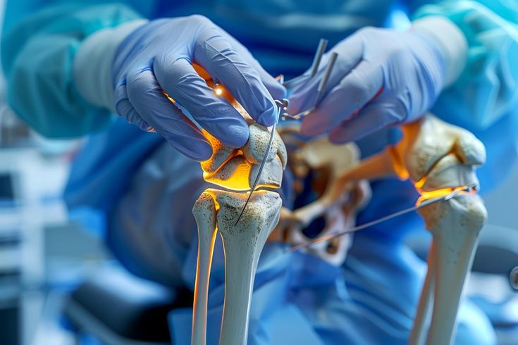
If the closed reduction surgery is not successful or if the hip dislocates again as the child starts to walk after spica removal it requires a major surgery. The hip joint is reduced by open method. Usually the shape of the femur bone needs to be changed by cutting the bone and fixing in desired angle with screw and plate. This method is commonly used in the age group of 1.5- 3 years.
Children who present late – after 3 years of age or so need additional procedure to alter the orientation or capacity of the acetabular socket to maintain the stability of hip joint.
Unfortunately, some hips do not continue to develop normally even after they stabilize. Further treatment is sometimes required.